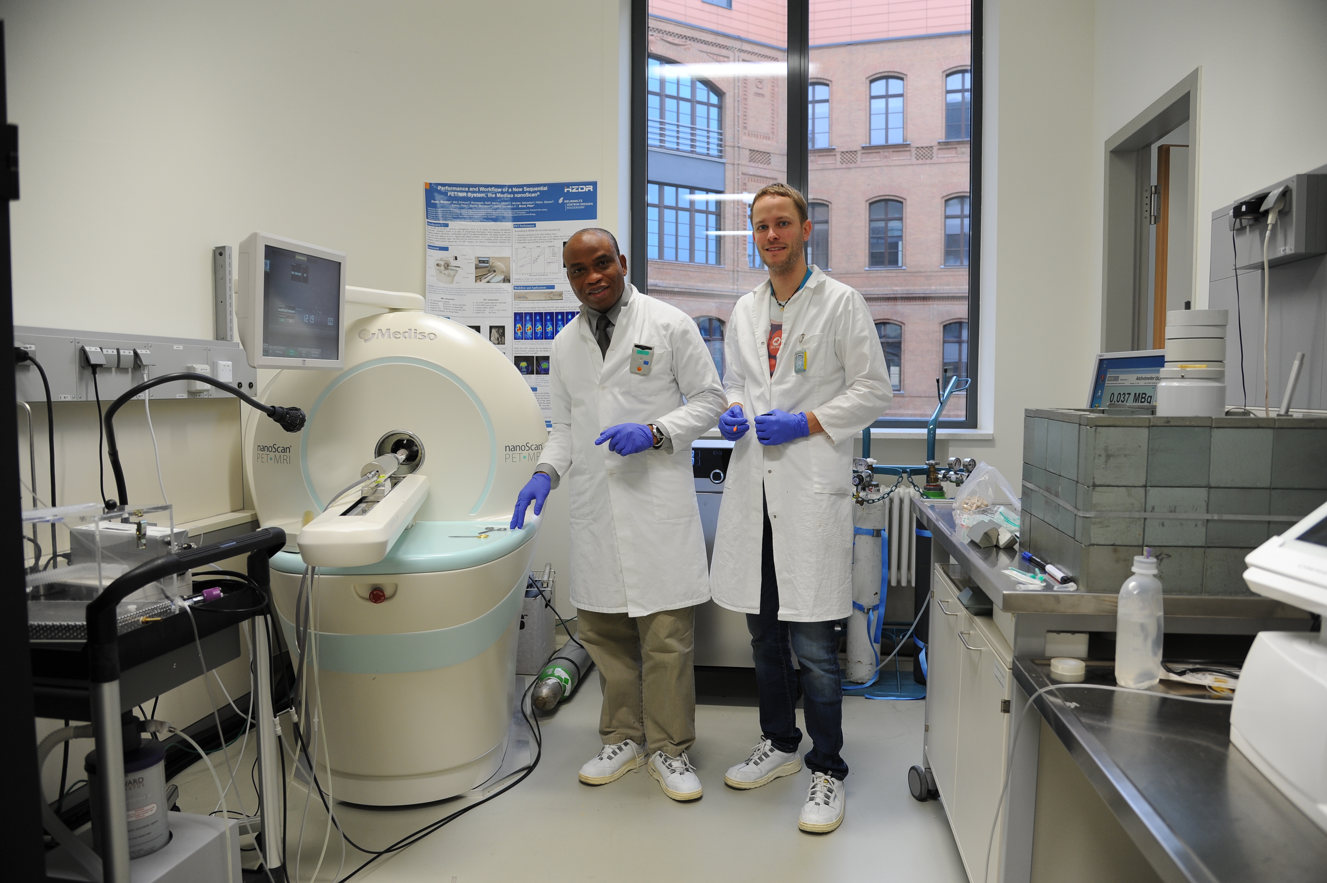
The race to create new super-computers with artificial intelligence that functions like human brains got a big boost with the latest breakthrough by Nigerian and German scientists. Scientists around the world are competing to unveil the mechanisms of brain function so that they can simulate it in new generation super-computers that work with artificial intelligence. Nigerian scientists are at the forefront of this international competition to create these future super-computers with artificial intelligence.
The major problem is that scientists do not really understand with precision the way the brain works and how differently it functions in male and female brains. This has been the subject of much scientific research and the breakthroughs keep coming. One of the intriguing problems relates to how information is taken from the time we see it to other areas of the brain for processing.
For example, what really happens in your brain when you see an apple on a tree? It happens that there are two different information super-highways in the brain. One called the “what” or ventral pathway, that runs from the back of the head towards the bottom side area and is responsible for recognizing the apple and its color. The other, information “super-highway” called the “where” or dorsal pathway, runs from the back of the head to the top middle area and locates where the apple is hanging relative to your position. Even though scientists have known this for a while, there was no precise technique to identify in which super-highway the information is being processed.
Developing such an imaging technique has been a mounting challenge. However, not anymore, as this may have been solved by this recent breakthrough. By applying a new technical innovation called functional positron emission tomography spectroscopy (fPETS) it was possible to precisely segregate information being processed in each brain information super-highway, either at the top of the brain called the cortical area or the bottom part of the brain called the subcortical region.
The investigators were surprised to demonstrate remarkable functional differences between male and female mice brains. In male mice brain, they showed that, the white day light activated the subcortical region in the dorsal pathway in the left half of the brain. On the other hand, the colored lights activated the cortical area in the ventral pathway of the same left half of the mice brain. It was a whole different picture in female mice, where the white light activated the cortical area in the ventral pathway in the right half of the brain.
While the colored light activated the subcortical region in the dorsal stream in the same right half of the brain. This first of its kind discovery is stunning both from the standpoint of gender differences in the brain and also from the insight which it provides scientists to model the mechanisms of brain function for artificial intelligence and other applications in health and disease. There are also benefits for diagnosis and clinical care of patients with memory deficits and strokes.
The social implications could be enormous, since the findings point to gender differences in learning mechanisms that could create different approaches in methods used for education. It also implies that male and female genders compliment each other rather than sameness.
We tried to find out the background of the work and how the Nigerian and German scientists were able to collaborate to make these major scientific discoveries. Scientists at the Neurocybernetic Flow laboratory, International Institutes of Advanced Research and Training, Chidicon Medical Center, Owerri led by Academician Prof Dr Philip Chidi Njemanze and their counterparts at the Helmholz Institute of Radiopharmacy, Dresden, at Leipzig University, Leipzig, Germany led by Prof Peter Brust have a long history of collaboration since 2015. They developed the new brain technique based on conventional positron emission tomography (PET) to which they applied advanced complex mathematical modeling called Fourier analysis.
The team was the first to apply the new ultrasensitive technique, which they called functional positron emission tomography spectroscopy (fPETS). It enhanced the computation of the index for brain metabolism measured by a radioactive dye. According to Prof Philip Njemanze, the lead author of the paper, “the innovation allowed us to pick up even very small differences in functional activation in different parts of the brain”.
Prof Brust who is a coauthor of the paper and head of the Helmholtz Institute laboratory in University of Leipzig, which is the largest Institute of Radiopharmacy in Europe, where the work was done, said “when our team with Njemanze’s group in 2017 discovered that color processing was different in male and female brain and published it in the American scientific journal PLoS ONE 12(7): e0179919. https://doi.org/10.1371/journal.pone.0179919, we could not offer much specifics of what was going on”.
Dr Mathias Kranze another coauthor added “when the proposal by Njemanze to zoom in on the brain pathways, we were pleased to collaborate and published the work on Fourier analysis in Forecasting 2019, 1(1), 121-142; https://doi.org/10.3390/forecast1010010 “. The authors intend to get scientists and doctors to use the technique, and just published it in a book by Intech Open Science, a global leading publisher at https://www.intechopen.com/online-first/application-of-fourier-analysis-of-cerebral-glucose-metabolism-in-color-induced-long-term-potentiati ”.
According to Njemanze, “the present work is one in a series that our neurocybernetic team here in Nigeria, focused to study the brain control systems. We hope we will be able to uncover the great secrets of the brain and get us closer to simulating human brain function with artificial intelligence”

